Friday, 28 August 2020
Thursday, 27 August 2020
Wednesday, 26 August 2020
Tuesday, 25 August 2020
Monday, 24 August 2020
Food Digestion
Process of food Digestion
Energy is required for different biological processes that take place in human body. Energy is obtained through food that we take into the body. These food contain nutrients such as carbohydrates, lipids and proteins. Carbohydrates, lipids and proteins are complex organic molecules that do not dissolve in water. These compounds cannot be absorbed into the human body. Therefore they should be broken down into small particles.
The process by which the complex organic compounds are converted into simple organic products to be absorbed into the human body is called digestion of food.
Food digestion takes place in two process namely mechanical and chemical processes
1. During mechanical process the physical nature of the food is altered,
E.g. :- Breaking down of food into small pieces by teeth inside mouth.
2.During chemical process, the insoluble complex compounds are broken down into simple molecules by the action of enzymes.
E.g. :- Starch is converted into maltose by salivary amylase (ptyalin) enzyme inside mouth.
There are some nutrients, that can be used by the body without any digestion, such as mineral salts, some vitamins, glucose, fructose and galactose. The organs involved in food digestion, are collectively called as digestive system.
Human digestive system
Human digestive system is a single tube, that runs from mouth to anus. According to the requirement, the structure has changed at different places, and the glands (salivary glands, pancreas, liver) that supply enzymes and other substances (bile) connect at different sites. The functions take place in the digestive system are food digestion, absorption of digested end products and removal of undigested materials from the body.

Digestion in the buccal cavity
Mouth opens the buccal cavity to the environment. It is surrounded by muscular lips at the bottom and top. The buccal cavity is made up of upper and lower jaws. Only the lower jaw can be moved. Teeth are present in both jaws. Buccal cavity is surrounded by cheeks. The tongue is attached to the floor of the buccal cavity. Three salivary glands are present in the buccal cavity. They secrete saliva and the tongue helps in identification of taste, mixing of food with saliva and swallowing.
The salivary amylase (ptyalin) enzyme, acts on starch in digestion of food. Starch will be partially digested into maltose. Digestion of food starts in the mouth.
There is a movable organ called epiglottis found just above the opening of trachea. When bolus is swallowed the epiglottis moves down to close the opening of trachea. Then bolus enters into oesophagus without entering into trachea.

Epiglottis helps to prevent entering food into the trachea. When food enters to pharynx, respiratory track is blocked by epiglottis. This prolong blockage of trachea may cause death. If the food is not removed instantly, the person may die due to blockage of respiratory tract.

The bolus passes through the oesophagus by peristaltic movements. As oesophagus is a muscular structure, due to contractions and relaxations of its wall the peristaltic movements appear as waves. These peristaltic movements provide the force to propel the bolus forward.
Then food is moved into stomach by peristaltic movements.
Digestion in the stomach
The stomach is a dilated sac like organ. Due to the peristaltic activity of muscles in the stomach wall the bolus is broken down and mixed well into a chyme. Several secretions ooze out into the stomach. It is collectively called the gastric juice.
The gastric juice contains mainly hydrochloric acid (HCl) and pepsin enzyme. HCl activates pepsin and pepsin starts the protein digestion to produce polypeptides. Renin present in infants causes coagulation of milk. Food retain in stomach for about three hours. Although the digested end products are not absorbed but some water, glucose and some drugs may absorb.
Chyme containing partially digested proteins, digested and undigested carbohydrates, undigested lipids, water, minerals and vitamins are released into the proximal part of small intestine, duodenum part by part.
When the stomach is empty, it continues to contract. When the stomach is empty for a longer period of time, the rate of contraction is also high. So it causes a pain. It gives a sense about hunger. Hunger is a signal that indicates the need of food.
Digestion in the small intestine
The chemical digestion of food mainly takes place in the small intestine. Pancreatic enzymes as well as intestinal enzymes involve in this digestion. The small intestine is about 7 m in length. The proximal part of the small intestine is C shaped and known as duodenum. The duct of the pancreas and the gall bladder opens into the duodenum via a single pore. Pancreatic juice is secreted into the duodenum through pancreatic duct. It contains three main enzymes. They are trypsin, amylase and lipase. The bile carried through the bile duct is added to it. Bile is produced in the liver and stored in the gall bladder.
Bile contains bile pigments, bile salts, bicarbonate ions and water
Due to mixing of bile with food at duodenum, the lipids in food are broken down into small droplets by the process called emulsification. Due to this action, enzymes get a greater surface area to act on lipid food.
Intestinal juice secreted by the wall of the intestine contains, maltase, sucrase, lactase, peptidase and mucus. Mucus lubricates food and then ease the passage of food materials along the gut. It protects the inner lining of gut wall. Proteins present in wall of stomach and intestine is protected by the protein digestive enzymes as there is a layer of mucus on the wall.
What happens to the end products of food digestion?
- Being a long tube
- Presence of circular folds in the inner wall
- Presence of finger like projections called villi in the circular folds
- Presence of microvilli in the epithelial cells of villi
- Thin epithelial lining on villi
- Villi are highly vascularised

The digestive end products given below are absorbed into the blood capillaries of villi.
- Amino acids
- Vitamins
- Mineral salts
- Monosaccharides
Fatty acids and glycerol formed by digestion of lipids are absorbed into lacteals. Finally they enter into blood circulatory system. When there is high amount of glucose in blood, they are converted into glycogen and stored in liver. In the same way when the concentration of glucose is decreased, glycogen breaks down to form glucose and is added to blood. The unabsorbed materials in small intestine are sent to the large intestine.
Processes in the large intestine

Length of the large intestine is about 1.5 m. It starts with caecum and ends up at anus. The dilated part of the large intestine is the rectum. The opening of it, is the anus. The materials entering into the large intestine contain a very small amount of nutrients. Mainly it contains undigested cellulose and water. A small blind ended tubular structure starts at the end of the caecum. It is known as the appendix. It is very small in humans and it may be infected and become swollen. This disease is known as appendicitis.
The main function of the large intestine is to absorb water from matter received from ileum. Thereby making it into semi solid. The matter enterd into large intestine is known as faecal matter. Faecal matter is yellow in colour due to bile pigments in it. Undigested food, microorganisms, epithelial cells and mucus are present in faecal matter.
When large intestine fills with faecal matter, it passes out from the rectum.
Saturday, 22 August 2020
Living tissues
Plant tissues
Meristematic tissues
The tissues with cells that divide actively by mitosis to produce new cells are called meristematic tissues. These cells are not differentiated. The growth of plants takes place due to activity of meristematic tissues.Features of meristematic tissues
This tissue consists of small sized living cells.No intercellular spaces or intercellular spaces are not prominent.There is a distinct nucleus in each cell.Absence of large central vacuole but small vacuoles may be present.Absence of chloroplasts.Large number of mitochondria are present.
Meristematic tissues are present in specific locations of the plant. They are of three types.

Apical meristems
Apical meristems are found in shoot apex, root apex and axillary buds. Plant increases its height due to the activity of this tissue.
Intercalary meristems
Intercalary meristems are found at nodes. The length of internode increases due to the activity of the above tissue. They are found in plants of grass family.
Lateral meristems
Lateral meristems are present laterally in the stem and roots of plant. They are found parallel to the longitudinal axis of plant. The diameter of the plant increases due to the activity of this tissue. Cambium tissue found in dicots is a lateral meristematic tissue.
Permanent tissues
According to the nature of the permanent tissues, it can be grouped into two.Simple Permanent tissues - One type of cells collected together
Complex Permanent tissues - Different types of cells collected together
Simple permanent tissues
The tissue is composed of similar cells. According to the shape of cell and the nature of cell wall, three types of simple permanent tissues as parenchyma, collenchyma and sclerenchyma can be identified in plants

Parenchyma
The tissue that forms the soft parts of the plant body is the parenchyma tissue. This is the most abundant tissue found in the plant.
Parenchyma tissue consists of living cells.
Cells are isodiametric (spherical) with a large central vacuole.
Nucleus is present peripherally in the cytoplasm.
The cell wall is thin and made up of cellulose.
Intercellular spaces are present.
Locations of parenchyma tissues
- Cortex and pith of plant stem
- Pith and cortex of roots
- Fleshy parts of fruits
- Seeds (endosperm)
- Leaves (mesophylls)
Collenchyma
Collenchyma tissue provides mechanical strength and support to the plant body. They are modified parenchyma cells.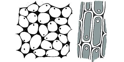
Features of collenchyma tissue
Collenchyma tissue consists of living cells.
Cells possess a cytoplasm, nucleus and central vacuole
Generally cells are elongated and polygonal in cross section.
The corners of the cell walls are thickened.
Intercellular spaces may present or may not present.
The collenchyma forms a cylindrical tissue inner to the epidermis of herbaceous stems. They are found in the veins of dicot leaves.
Sclerenchyma
Sclerenchyma tissue helps in providing mechanical strength and support to the plant body. This tissue has two types of cells as sclereids and sclerenchyma fibres.
Features of sclerenchyma tissue
Fibres present in xylem are called as xylem fibres and in phloem as phloem fibres. Other than above, coconut fibres, agave fibres and cotton wool are made up of fibres (sclerenchyma)Sclerenchyma tissue consists of dead cells.Lignin is deposited on the cellulose cell wall.Cell wall is evenly thickened and forms a central lumen.Cells are tightly packed. Therefore, no intercellular spaces.
Selereids are found in endocarp of coconut, Kaduru and mango fruits, the pericarp of guava fruit and in pear fruit and seed coat of coffee and dates.
Animal tissues
The animal body is also made up of different types of cells. Example :- The human body is made up of about 210 different types of cells. There are groups of cells with common origin to perform a particular function in the multicellular animal body. Main types of animal tissues are given below.
- Epithelial tissue
- Connective tissue
- Muscle tissue
- Nervous tissue

Epithelial tissue
This is the tissue that lines up the free surfaces (internal and external) of the vertebrate body. Some of them are composed of single layer of cells and the others are with several cell layers.
The epithelial tissue is classified according to the shape of the cell and the number of cell layers.The cells are placed on a basement membraneThe cells are tightly packedA nerve supply is present within the tissue but there is no blood supply
Examples for several locations of epithelial tissues are given below.
Functions of epithelial tissue
- Wall of blood capillaries
- Thyroid gland
- Lining of nasal cavity
- Wall of urinary bladder
- Skin (Epidermis)
Lining up of free surfaces and protection - Protects the internal organs from pressure, friction and microbes
Absorptive function - The epithelium of digestive tract absorbs digestive end products
Perception of stimuli - The epithelium of tongue and nose, detect taste and smell senses
Secretory function - Secretion of mucous by the lining epithelium of respiratory tract
Filtering function - Epithelium of Bowman's capsule in nephrons, filters blood
Connective tissue

Connective tissue is composed of different types of cells and fibres. These cells and fibres are embedded in a large matrix. Most connective tissues possess nerve and blood supply.
The connective tissues provide connection between tissues and organs and provide support too.
E.g. :- Blood tissue, Bone tissue
Blood tissue
Blood is a special connective tissue. The speciality is that the matrix (plasma) is not secreted by the blood cells. Blood tissue helps in maintaining proper connection between organs and tissue of the human body.
Blood is composed of a fluid matrix. Matrix is called plasmaThe matrix contains cells called red blood cells (erythrocytes) and white blood cells (leucocytes) and cellular fragments called plateletsFibres are not found always but during blood clotting they appear
Functions of blood tissue
- Transportation of materials - Respiratory gases, nutrients, excretory materials and hormones are transported to the relevant organs
- Protection - White blood cells destroy foreign bodies (Microbes) by phagocytosis and by producing antibodies
- Maintenance of homeostasis

Muscle tissue
Muscle tissue is one of the main tissues that makes up the human body. Muscle tissue is made up of muscle cells or muscle fibres. These muscle fibres possess contraction and relaxation ability. Not like epithelial tissue, the muscle tissue possesses a good blood supply. Therefore muscle tissue receives oxygen and nutrients at a high rate. Muscle tissue acts as one of the effectors in responding in coordination.Muscle tissue is of three types,
- Smooth muscle tissue
- Skeletal muscle tissue
- Cardiac muscle tissue
Smooth muscle tissue
Smooth muscle tissue is made up of smooth muscle cells. This tissue is found in the walls of organs with cavities.Example :- Walls of digestive tract, uterus, blood vessels and bladder

- These cells are spindle shaped. The cells are unbranched
- These cells have one nucleus at the centre. No striations
- These cells do not become fatigue quickly. They are controlled involuntarily
Skeletal muscle tissue
Skeletal muscle tissue is made up of skeletal muscle fibres. These are mostly associated with skeletal system. The skeletal muscles help in locomotion and movements of chordates.
E.g. :- Bicep muscle, Tricep muscle, Muscles in leg, Facial muscles
- Skeletal muscle fibres are long, cylindrical, unbranched cells.
- They are multinucleate cells with striations. The nuclei present peripherally, and many mitochondria are present.
- These cells are voluntarily controlled and become fatigue easily.
Cardiac muscle tissue
Cardiac muscle tissue is made up of cardiac muscle cells. It is exclusively found in the vertebrate heart.
- Cardiac muscle cells are uninucleate, striated and short cells
- Intercalated discs are present among cells
- They never become fatigue. They contract rhythmically
- They are involuntarily controlled
Nervous tissue

It is an important tissue found in chordates body. The structural unit of nervous tissue is nerve cell or neuron. Neurons are specialised to transmit impulses.
Features of neuron
Functions of neuronsNeuron is composed of two parts. They are cell body and nerve fibres.Nucleus, mitochondria, golgi body and endoplasmic reticulum are found in the cell body.Axon arises from the cell body as a single process. The axon transmits impulses away from the cell body.Slender process called dendrites receive stimuli and transmit impulses to the cell body.Most of the axons in chordates are myelinated. Myelin sheath is not continuous and the interrupted places are known as nodes of Ranvier. The myelin sheath increases the speed of transmission of impulses.
The function of the neuron is to receive the information from the receptors (eye, ear, nose, tongue, skin) or another neuron and to transmit them to the effector (muscles) or to another neuron. According to the function of the neuron, they can be divided into three types as follows.
- Sensory neuron
- Inter neuron
- Motor neuron
functions of the plant and animal cell
Concept of the cell
The cell is the smallest structural unit of the organization of the living body. The organisms composed of a single cell are called unicellular organisms and those of many cells are called multicellular organisms. Cells perform different functions in the body.
For example - The transportation of oxygen is done by red blood cells. Transmission of impulses is done by neurons. Accordingly, the smallest bio unit that is adapted to perform a particular function is the cell. So it is clear that the structural and functional unit of life is the cell. The cells differ from one another from their shape, size and function. Except few occasions, mostly cells are not visible to the naked eye. Therefore they have to be observed using the light microscope.
Typical cell The small structures present within the cell to perform different functions are known as organelles.The types of organelles and the number of them differ according to the function performed by the cell. The cell prepared by including all the organelles is known as the typical cell. In the living world such cells do not exist. But cells with a certain number of organelles of the typical cell can be found in living organisms.

All animal cells are covered by a plasma membrane or a cell membrane. It is a live semi permeable membrane as well as a selective permeable membrane. There is a centralized nucleus in an animal cell. The cytoplasm is a gelatinous material. The outer covering of the plant cell is the cell wall . It is made up of cellulose. Inner to the cell wall is the plasma membrane. At the center of plant cell is a large vacuole. Generally there are no such vacuoles in animal cells. Animal cells as well as plant cells possess different organelles that perform different functions. Most of the above organelles cannot be observed through the light microscope. Therefore the electron microscope should be used. Below are the typical plant and animal cells created based on electron microscopic information.


Subscribe to:
Posts (Atom)