Plant tissues
Meristematic tissues
The tissues with cells that divide actively by mitosis to produce new cells are called meristematic tissues. These cells are not differentiated. The growth of plants takes place due to activity of meristematic tissues.Features of meristematic tissues
This tissue consists of small sized living cells.No intercellular spaces or intercellular spaces are not prominent.There is a distinct nucleus in each cell.Absence of large central vacuole but small vacuoles may be present.Absence of chloroplasts.Large number of mitochondria are present.
Meristematic tissues are present in specific locations of the plant. They are of three types.

Apical meristems
Apical meristems are found in shoot apex, root apex and axillary buds. Plant increases its height due to the activity of this tissue.
Intercalary meristems
Intercalary meristems are found at nodes. The length of internode increases due to the activity of the above tissue. They are found in plants of grass family.
Lateral meristems
Lateral meristems are present laterally in the stem and roots of plant. They are found parallel to the longitudinal axis of plant. The diameter of the plant increases due to the activity of this tissue. Cambium tissue found in dicots is a lateral meristematic tissue.
Permanent tissues
According to the nature of the permanent tissues, it can be grouped into two.Simple Permanent tissues - One type of cells collected together
Complex Permanent tissues - Different types of cells collected together
Simple permanent tissues
The tissue is composed of similar cells. According to the shape of cell and the nature of cell wall, three types of simple permanent tissues as parenchyma, collenchyma and sclerenchyma can be identified in plants

Parenchyma
The tissue that forms the soft parts of the plant body is the parenchyma tissue. This is the most abundant tissue found in the plant.
Parenchyma tissue consists of living cells.
Cells are isodiametric (spherical) with a large central vacuole.
Nucleus is present peripherally in the cytoplasm.
The cell wall is thin and made up of cellulose.
Intercellular spaces are present.
- Cortex and pith of plant stem
- Pith and cortex of roots
- Fleshy parts of fruits
- Seeds (endosperm)
- Leaves (mesophylls)
Collenchyma
Collenchyma tissue provides mechanical strength and support to the plant body. They are modified parenchyma cells.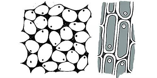
Features of collenchyma tissue
Collenchyma tissue consists of living cells.
Cells possess a cytoplasm, nucleus and central vacuole
Generally cells are elongated and polygonal in cross section.
The corners of the cell walls are thickened.
Intercellular spaces may present or may not present.
The collenchyma forms a cylindrical tissue inner to the epidermis of herbaceous stems. They are found in the veins of dicot leaves.
Sclerenchyma
Sclerenchyma tissue helps in providing mechanical strength and support to the plant body. This tissue has two types of cells as sclereids and sclerenchyma fibres.
Features of sclerenchyma tissue
Fibres present in xylem are called as xylem fibres and in phloem as phloem fibres. Other than above, coconut fibres, agave fibres and cotton wool are made up of fibres (sclerenchyma)Sclerenchyma tissue consists of dead cells.Lignin is deposited on the cellulose cell wall.Cell wall is evenly thickened and forms a central lumen.Cells are tightly packed. Therefore, no intercellular spaces.
Selereids are found in endocarp of coconut, Kaduru and mango fruits, the pericarp of guava fruit and in pear fruit and seed coat of coffee and dates.
Animal tissues
The animal body is also made up of different types of cells. Example :- The human body is made up of about 210 different types of cells. There are groups of cells with common origin to perform a particular function in the multicellular animal body. Main types of animal tissues are given below.
- Epithelial tissue
- Connective tissue
- Muscle tissue
- Nervous tissue

Epithelial tissue
This is the tissue that lines up the free surfaces (internal and external) of the vertebrate body. Some of them are composed of single layer of cells and the others are with several cell layers.
The epithelial tissue is classified according to the shape of the cell and the number of cell layers.The cells are placed on a basement membraneThe cells are tightly packedA nerve supply is present within the tissue but there is no blood supply
Examples for several locations of epithelial tissues are given below.
Functions of epithelial tissue
- Wall of blood capillaries
- Thyroid gland
- Lining of nasal cavity
- Wall of urinary bladder
- Skin (Epidermis)
Lining up of free surfaces and protection - Protects the internal organs from pressure, friction and microbes
Absorptive function - The epithelium of digestive tract absorbs digestive end products
Perception of stimuli - The epithelium of tongue and nose, detect taste and smell senses
Secretory function - Secretion of mucous by the lining epithelium of respiratory tract
Filtering function - Epithelium of Bowman's capsule in nephrons, filters blood
Connective tissue

Connective tissue is composed of different types of cells and fibres. These cells and fibres are embedded in a large matrix. Most connective tissues possess nerve and blood supply.
The connective tissues provide connection between tissues and organs and provide support too.
E.g. :- Blood tissue, Bone tissue
Blood tissue
Blood is a special connective tissue. The speciality is that the matrix (plasma) is not secreted by the blood cells. Blood tissue helps in maintaining proper connection between organs and tissue of the human body.
Blood is composed of a fluid matrix. Matrix is called plasmaThe matrix contains cells called red blood cells (erythrocytes) and white blood cells (leucocytes) and cellular fragments called plateletsFibres are not found always but during blood clotting they appear
- Transportation of materials - Respiratory gases, nutrients, excretory materials and hormones are transported to the relevant organs
- Protection - White blood cells destroy foreign bodies (Microbes) by phagocytosis and by producing antibodies
- Maintenance of homeostasis

Muscle tissue
Muscle tissue is one of the main tissues that makes up the human body. Muscle tissue is made up of muscle cells or muscle fibres. These muscle fibres possess contraction and relaxation ability. Not like epithelial tissue, the muscle tissue possesses a good blood supply. Therefore muscle tissue receives oxygen and nutrients at a high rate. Muscle tissue acts as one of the effectors in responding in coordination.
- Smooth muscle tissue
- Skeletal muscle tissue
- Cardiac muscle tissue
Smooth muscle tissue
Smooth muscle tissue is made up of smooth muscle cells. This tissue is found in the walls of organs with cavities.Example :- Walls of digestive tract, uterus, blood vessels and bladder

- These cells are spindle shaped. The cells are unbranched
- These cells have one nucleus at the centre. No striations
- These cells do not become fatigue quickly. They are controlled involuntarily
Skeletal muscle tissue
Skeletal muscle tissue is made up of skeletal muscle fibres. These are mostly associated with skeletal system. The skeletal muscles help in locomotion and movements of chordates.
E.g. :- Bicep muscle, Tricep muscle, Muscles in leg, Facial muscles
- Skeletal muscle fibres are long, cylindrical, unbranched cells.
- They are multinucleate cells with striations. The nuclei present peripherally, and many mitochondria are present.
- These cells are voluntarily controlled and become fatigue easily.
Cardiac muscle tissue
Cardiac muscle tissue is made up of cardiac muscle cells. It is exclusively found in the vertebrate heart.
- Cardiac muscle cells are uninucleate, striated and short cells
- Intercalated discs are present among cells
- They never become fatigue. They contract rhythmically
- They are involuntarily controlled
Nervous tissue

It is an important tissue found in chordates body. The structural unit of nervous tissue is nerve cell or neuron. Neurons are specialised to transmit impulses.
Features of neuron
Functions of neuronsNeuron is composed of two parts. They are cell body and nerve fibres.Nucleus, mitochondria, golgi body and endoplasmic reticulum are found in the cell body.Axon arises from the cell body as a single process. The axon transmits impulses away from the cell body.Slender process called dendrites receive stimuli and transmit impulses to the cell body.Most of the axons in chordates are myelinated. Myelin sheath is not continuous and the interrupted places are known as nodes of Ranvier. The myelin sheath increases the speed of transmission of impulses.
- Sensory neuron
- Inter neuron
- Motor neuron